Click on the following link for the main website: Marine Sponges from Hawaii
Marine sponges were once thought to be one of the simplest multicellular organisms of the metazoans. Instead, these organisms are actually highly complex and ecologically important to the reef ecosystem, especially in the Caribbean. Diaz and Rützler (2001) highlighted at least six important functional roles sponges play in Caribbean coral reefs: primary production and nitrification through complex symbiosis; chemical and physical adaptation for successful space competition; capability to impact the carbonate framework through calcification, cementation and bio-erosion; and the potential to alter the water column and its processes through high water filtering capabilities. As of August 31, 2011 the number of taxonomically recognized species of Porifera is 8,553, the vast majority of which (83%) are Demospongiae (Van Soest et al. 2012). In the same study, Van Soest et al. (2012) predicted that at the end of the present century the number of known Porifera species will have risen to at least 12,000. New species are awaiting to be discovered by scientists all over the world. Sponges are widely distributed in aquatic systems and successfully in habit hard- and soft-bottom communities, from tropical to polar latitudes, littoral to abyssal habitats, and fresh- to saltwater, being one of the major groups in both biomass and number of species in hard-bottom communities (Sarà & Vacelet, 1973).
Because of its isolation, the Hawaiian archipelago allows scientists to study an array of endemic species; marine sponges being one of them. In this blog you will find in situ pictures, descriptions and spicule pictures of marine sponges found on the east coast of the Island of Hawaii.
*I am not a marine sponge taxonomist. Marine sponges will be properly identified using their spicules by Dr. de Voogd, a leader in marine sponge identification. Sponge spicule pictures were taken using the Scanning Electron Microscope, which is located in the Marine Science building at the University of Hawaii at Hilo.
Site Locations:
Sponges were collected from Leliwii Beach Park or Richardson’s beach park using scuba diving equipment or from Chalks tide pool using snorkel.

Collection Site Map
References:
Diaz MC, Rützler K (2001) Sponges: An essential component of Caribbean coral reefs. Bulletin of marine Science, 69: 535-546
Sarà, M & Vacelet, J (1973) Ecologie des Démosponges. In: Grassé, P.P. (Ed.), Spongiaires. Masson, Paris: Masson et Cie. 462-576
Van Soest Rob WM, Boury-Esnault N, Vacelet J, Dohrmann M, Erpenbeck D, de Voogd NJ, Santodomingo N, Vanhoorne B, Kelly M and Hooper NAJ (2012) Global Diversity of Sponges (Porifera). PLoS ONE 7: 1-23
———————————————————————————————————–
Marine Sponge: Laboratory ID – HS0001: Clathrina sp. (Calcarea)

Clathrina sp. : in situ

Clathrina sp. : in situ size reference (in)

Clathrina sp. : Tissue Analysis

Clathrina sp. : Tissue Analysis

Clathrina sp. : Tissue Analysis

Clathrina sp. : Spicule Diversity Analysis

Clathrina sp. : Spicule Diversity Analysis

Clathrina sp. : Spicule Diversity Analysis
Description:
This bright neon yellow sponge (Clathrina sp.) has been observed growing/attached on dead Pocillopora meandrina (cauliflower coral) and Pocillopora eydouxi (antler coral). Sponge tends to grow in ‘patches’, not as a large individual sponge. It would be really interesting to see whether the sponge ‘patches’ are clones of each other or separate individuals of the same species. It is also really soft and ‘spongy’ to the touch. There was no noticeable irritation of the skin or any other post-handle ling symptoms related to touching this sponge. I have only observed this sponge while scuba diving, never in any tide pool environment that I have surveyed, ie: Onekahakaha beach park or chalks. Picture was taken at a depth of about 3 meters, but I have seen it at deeper depths, ranging from 1-20 meters. Ostia are clearly visible, the osculum is tougher to identify. Predators, if any, have not been observed or identified.
———————————————————————————————————–
Marine Sponge: Laboratory ID – HS0002: Spirastrella keaukahu

S. keaukahu : in situ

S. keaukahu : insitu dirty’ orange sponge size (cm)

S. keaukahu : Surface size reference (cm)

S. Keaukahu : Spicule Diversity Analysis

S. keaukahu : Spicule Diversity Analysis

S. Keaukahu : Spicule Diversity Analysis

S. keaukahu : Spicule Diversity Analysis

S. keaukahu : Spicule Diversity Analysis

S. keaukahu : Spicule Diversity Analysis

S. keaukahu : Sponge Tissue Analysis

S. Keaukahu : Sponge Tissue Analysis
Description:
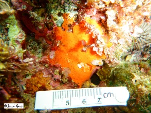
I have observed this ‘dirty’ rugged looking orange sponge (Spirastrella keaukahu) living in between crevices in shallow environments. Have yet to see it while scuba diving. Its surface appears rough, but it is actually quite smooth and easily torn. It is also spongy to the touch, did not develop any irritations or other symptoms after handling this sponge. Relatively large osculums (<1cm) are visible, with really small ostia. I have also observed a grayish sponge growing near this sponge.
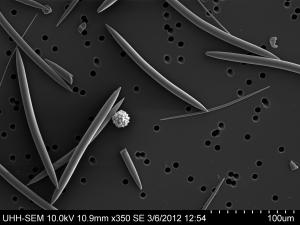
S.E.M analysis: I was able to identify at least twelve spicules, some might be the same type of spicule but just vary in size. Besides spicule pictures, I also took pictures of the sponge tissue so that we can analyze how the spicules might be interconnected within the sponge tissue.
———————————————————————————————————–
Marine Sponge: Laboratory ID – HS0003: sample too small for identification

HS0003: insitu Baby Blue Sponge

HS0003: insitu Baby Blue Sponge

HS0003: Sponge Tissue Analysis

HS0003: Sponge Tissue Analysis

HS0003: Spicule Diversity Analysis

HS0003: Spicule Diversity Analysis

HS0003: Spicule Diversity Analysis
Description:
I have only seen this sponge once. I managed to collect a small sample so that I can perform spicule and tissue analysis using the scanning electron microscope. It was encrusted on rock substrate.
———————————————————————————————————–
Marine Sponge: Laboratory ID – HS0004: Unidentified Bright red/orange Sponge

HS0004: Insitu size reference (in)

HS0004: Surface size reference (cm)

HS0004: Tissue Analysis

HS0004: Tissue Analysis

HS0004: Tissue Analysis

HS0004: Spicule Diversity Analysis

HS0004: Spicule Diversity Analysis

HS0004: Spicule Diversity Analysis

HS0004: Spicule Diversity Analysis
Description:
This bright red orange encrusting sponge was collected from Chalks. It is thin and slimy to the touch, in situ and topside. Sponge released some sort of mucous that covered the whole piece of sponge collected. It was collected from a depth of about 1.5 meters.
———————————————————————————————————–
Marine Sponge: Laboratory ID – HS0005: Haliclona sp.

HS0005: Blue Green Sponge

HS0005: Insitu size reference (in)

HS0005: Surface size reference (cm)

HS0005: Tissue Analysis

HS0005: Tissue Analysis

HS0005: Tissue Analysis

HS0005: Tissue Analysis

HS0005: Spicule Diversity Analysis

HS0005: Spicule Diversity Analysis

HS0005: Spicule Diversity Analysis

HS0005: Spicule Diversity Analysis
Description:
This blue green sponge (Haliclona sp.) was collected from chalks at a depth of about 1.5 meters. This sponge is really hard in texture, really tough. No irritation post handling the sponge. Osculums and ostia are clearly visible, internal structure can also be contemplated by looking through a large enough osculum. Although this sponge is encrusted onto the substrate, it is not as thing as most of the other sponges I have encountered, it is relatively thicker. I have not observed any predators that might be feeding on this sponge. *this sponge might be the same species as HS0006, HS0007 and HS0008. It might be that they just differ in color, although, after spicule diversity analysis, it is obvious that there are spicules that were found in HS0005 and not in the others. Color might also be a difference in sex between species. Proper genetic analysis needs to be perform in order to confirm my hypothesis.
———————————————————————————————————–
Marine Sponge: L6aboratory ID – HS0006: Haliclona sp 2.

Haliclona sp. 2 : Purple Sponge

Haliclona sp. 2 : in situ size reference (cm)

Haliclona sp. 2 : Surface size reference (cm)

Haliclona sp. 2 : Spicule Diversity Analysis

Haliclona sp. 2 : Spicule Diversity Analysis

Haliclona sp. 2 : Spicule Diversity Analysis
Description:
This purple sponge (Haliclona sp. 2) was collected from chalks from a depth of about 1.5 meters. It has a strong resemblance as HS0005. It was also a thick encrusted sponge, that has a sturdy texture and really tough. It is not easily torn from the substrate. One of the differences between this sponge and HS0005 is that this sponge does not have as many osculum present. Tissue looks similar. It is important to note that the preliminary sponge spicule analysis showed that they have a slight difference in spicule diversity.
———————————————————————————————————–
Marine Sponge: Laboratory ID – HS0007: Haliclona sp 2.

Halicona sp. 2 : Bright Violet Sponge

Haliclona sp. 2 : In situ size reference (in)

Haliclona sp. 2 : Tissue Analysis

Haliclona sp. 2 : Tissue Analysis

Haliclona sp. 2 : Spicule Diversity Analysis

Haliclona sp. 2 : Spicule Diversity Analysis
Description:
This bright violet sponge (Haliclona sp.) was collected from chalks at a depth of about 1.5 meters. This sponge also resembles HS0005, HS0006 and HS0008, in morphology and appearance. This sponge is bright violet instead of purple. This sponge had a significant percent of it covered in algae, something that I had not observed before in any of the other sponges that I have seen. Osculum and ostia are clearly visible in this sponge, there was also no irritation to the skin post handling of this sponge.
———————————————————————————————————–
Marine Sponge: Laboratory ID – HS0008: Haliclona sp.

HS0008: Purple Sponge “castle-like”

HS0008: Insitu size reference (cm)

HS0008: Outside size reference (cm)

HS0008: Spicule Diversity Analysis

HS0008: Spicule Diversity Analysis

HS0008: Spicule Diversity Analysis
Description: Haliclona sp.
———————————————————————————————————–
Marine Sponge: Laboratory ID – HS0009: Iotrochota protea

HS0009: Black Sponge

HS0009: Insitu size reference (cm)

HS0009: Surface size reference

HS0009: Tissue Analysis

HS0009: Tissue Analysis

HS0009: Tissue Analysis

HS0009: Tissue Analysis

HS0009: Tissue Analysis

HS0009: Spicule Diversity Analysis

HS0009: Spicule Diversity Analysis

HS0009: Spicule Diversity Analysis

HS0009: Spicule Diversity Analysis
Description:
This black sponge (Iotrochota protea) was collected from chalks at a depth of about 1 meter. It forms irregular encrusting thick round lobes. Prominent osculum can be seen with really small ostias. This sponge is smooth to the touch, it releases some sort of mucous once it is being handled. It turned 80% ethanol solution dark red. No irritation was noticed post handling of this sponge.
———————————————————————————————————–
Marine Sponge: Laboratory ID – HS0010: Clathria sp.

HS0010: Encrusted dull orange sponge

HS0010: Insitu size reference

HS0010: Spicule Diversity Analysis

HS0010: Spicule Diversity Analysis

HS0010: Spicule Diversity Analysis

HS0010: Spicule Diversity Analysis

HS0010: Spicule Diversity Analysis
Description:
This really small paper thin encrusted sponge (Clathria sp.) was collected off Leleiwii beach park using scuba diving equipment from a depth of about 7 meters. Enough tissue was taken just for a spicule diversity analysis, a tissue analysis was not performed. I have seen this sponge encrusted only in small rocks. Osculum was barely visible, ostia were not.
———————————————————————————————————–
Marine Sponge: Laboratory ID – HS0011: Haliclona sp. 1

HS0011: Dirty yellow sponge

HS0011: Surface size reference (cm)

HS0011: Tissue Analysis

HS0011: Tissue Analysis

HS0011: Spicule Diversity Analysis

HS0011: Tissue Analysis

HS0011: Spicule Diversity Analysis

HS0011: Spicule Diversity Analysis

HS0011: Spicule Diversity Analysis
Description:
This yellow colored Haliclona sp. 1 was collected from an overhang/cave, and it was not seen out in the open reef or exposed to the sunlight.
———————————————————————————————————–
Marine Sponge: Laboratory ID – DS0012: Unidentified

DS0012: Tissue Analysis

DS0012: Tissue Analysis

DS0012: Tissue Analysis

DS0012: Tissue Analysis

DS0012: Tissue Analysis

DS0012: Tissue Analysis

DS0012: Spicule Diversity Analysis

DS0012: Spicule Diversity Analysis

DS0012: Spicule Diversity Analysis
Description:
Mesophotic reef sponge (Tethya aff communis), collected from the Au’au Channel off Maui using the NOAA HURL PISCES IV and PISCES V submersibles from February 26 – March 10, 2011 (Parrish/Rooney) at a depth of 121 meters.
————————————————————————————————————–
Marine Sponge: Laboratory ID – DS0013: Unidentified

DS0013: Tissue Analysis

DS0013: Tissue Analysis

DS0013: Tissue Analysis

DS0013: Tissue Analysis

DS0013: Tissue Analysis

DS0013: Tissue Analysis

DS0013: Spicule Diversity Analysis

DS0013: Spicule Diversity Analysis

DS0013: Spicule Diversity Analysis

DS0013: Spicule Diversity Analysis

DS0013: Spicule Diversity Analysis
Description:
Mesophotic reef sponge (Dactylospongia aff metachromia), collected from the Au’au Channel off Maui using the NOAA HURL PISCES IV and PISCES V submersibles from February 26 – March 10, 2011 (Parrish/Rooney) at a depth of 108 meters.
———————————————————————————————————–
Marine Sponge: Laboratory ID – DS0014: Unidentified

DS0014: Tissue Analysis

DS0014: Tissue Analysis

DS0014: Tissue Analysis

DS0014: Tissue Analysis

DS0014: Tissue Analysis

DS0014: Spicule Diversity Analysis

DS0014: Spicule Diversity Analysis

DS0014: Spicule Diversity Analysis

DS0014: Spicule Diversity Analysis

DS0014: Spicule Diversity Analysis
Description:
Mesophotic reef sponge (unidentified), collected from the Au’au Channel off Maui using the NOAA HURL PISCES IV and PISCES V submersibles from February 26 – March 10, 2011 (Parrish/Rooney) at a depth of 108 meters.
———————————————————————————————————–
Marine Sponge: Laboratory ID – DS0015: Deep Sponge (see description)
Description:
Mesophotic reef marine sponge (unidentified), collected from the Au’au Channel off Maui using the NOAA HURL PISCES IV and PISCES V submersibles from February 26 – March 10, 2011 (Parrish/Rooney) at a depth of 85 meters.
———————————————————————————————————–
Marine Sponge: Laboratory ID – DS0016: Deep Sponge (see description)
Description:
Mesophotic reef marine sponge (unidentified), collected from the Au’au Channel off Maui using the NOAA HURL PISCES IV and PISCES V submersibles from February 26 – March 10, 2011 (Parrish/Rooney) at a depth of 85 meters.
———————————————————————————————————–
Marine Sponge: Laboratory ID – HS0017: Charcoal sponge

HS0017: Insitu Charcoal color sponge

HS0017: Charcoal color sponge

HS0017: Tissue Analysis

HS0017: Tissue Analysis

HS0017: Tissue Analysis

HS0017: Tissue Analysis

HS0017: Tissue Analysis

HS0017: Tissue Analysis

HS0017: Spicule Diversity Analysis

HS0017: Spicule Diversity Analysis

HS0017: Spicule Diversity Analysis

HS0017: Spicule Diversity Analysis

HS0017: Spicule Diversity Analysis

HS0017: Spicule Diversity Analysis

HS0017: Spicule Diversity Analysis

HS0017: Spicule Diversity Analysis

HS0017: Spicule Diversity Analysis

HS0017: Spicule Diversity Analysis

HS0017: Spicule Diversity Analysis

HS0017: Spicule Diversity Analysis

HS0017: Spicule Diversity Analysis

HS0017: Spicule Diversity Analysis

HS0017: Spicule Diversity Analysis

HS0017: Spicule Diversity Analysis

HS0017: Spicule Diversity Analysis

HS0017: Spicule Diversity Analysis

HS0017: Spicule Diversity Analysis
Description:
———————————————————————————————————–
Marine Sponge: Laboratory ID – HS0018: Lissodendoryx schmidti

HS0018: Orange Sponge

HS0018: Insitu size reference (cm)

HS0018: Surface Size Reference (cm)

HS0018: Tissue Analysis

HS0018: Tissue Analysis

HS0018: Tissue Analysis

HS0018: Tissue Analysis

HS0018: Tissue Analysis

HS0018: Spicule Diversity Analysis

HS0018: Spicule Diversity Analysis

HS0018: Spicule Diversity Analysis

HS0018: Spicule Diversity Analysis

HS0018: Spicule Diversity Analysis
Description:
Read Full Post »


